CHD1A (4B4C) Materials & Methods |
Entry clone source: MGC (IMAGE) |
Entry clone accession: IMAGE:40125685 |
SGC Construct ID: CHD1A-c107 |
Vector: pNIC28-Bsa4. Details [PDF ]; Sequence [ FASTA ] or [ GenBank ]. |
DNA sequence:
ATGCACCATCATCATCATCATTCTTC
TGGTGTAGATCTGGGTACCGAGAACC
TGTACTTCCAATCCATGCCTCGGGAG
AATATTAAAGGATTTAGTGATGCAGA
AATTAGGCGGTTTATCAAGAGCTATA
AGAAATTTGGTGGTCCTCTGGAAAGA
TTAGATGCAATTGCTCGAGATGCTGA
GTTAGTTGATAAGTCAGAAACAGACC
TTAGACGACTGGGAGAATTGGTACAT
AATGGTTGCATTAAAGCATTAAAGGA
TAGTTCTTCAGGAACAGAACGAACAG
GTGGTAGACTCGGAAAAGTGAAGGGT
CCAACATTCCGAATATCAGGAGTACA
GGTGAATGCCAAACTAGTCATCTCCC
ATGAAGAAGAATTAATACCTTTGCAC
AAATCCATTCCTTCTGATCCAGAAGA
AAGAAAGCAGTATACTATCCCATGCC
ACACAAAGGCAGCTCATTTTGATATA
GACTGGGGCAAAGAAGATGATTCCAA
TTTGTTAATTGGCATCTATGAATATG
GATATGGAAGCTGGGAAATGATTAAA
ATGGATCCTGACCTCAGTCTAACACA
CAAGATTCTTCCAGATGATCCCGATA
AAAAACCACAAGCAAAACAGTTGCAG
ACCCGTGCAGACTACCTCATCAAATT
ACTTAGTAGAGATCTTGCAAAAAAAG
AAGCTCTTTCTGGTGCGGGATGA
|
Final protein sequence (His6 affinity Tag sequence in lowercase):
mhhhhhhssgvdlgtenlyfq^smPR
ENIKGFSDAEIRRFIKSYKKFGGPLE
RLDAIARDAELVDKSETDLRRLGELV
HNGCIKALKDSSSGTERTGGRLGKVK
GPTFRISGVQVNAKLVISHEEELIPL
HKSIPSDPEERKQYTIPCHTKAAHFD
IDWGKEDDSNLLIGIYEYGYGSWEMI
KMDPDLSLTHKILPDDPDKKPQAKQL
QTRADYLIKLLSRDLAKKEALSGAG
^ TEV protease recognition site
|
Tags and additions: Cleavable N-terminal His6 tag |
Host: BL21(DE3)-R3-pRARE2. |
Cell Growth and Induction: The expression plasmid was transformed into the host strain and plated on LB-agar containing 50 µg/ml kanamycin and 34 µg/ml chloramphenicol. Several colonies were combined to inoculate a 1ml culture in TB (+ 50 µg/ml kanamycin, 34 µg/ml chloramphenicol). The culture was grown overnight, glycerol was added to 15% v/v (from a 60% stock), and the resulting glycerol stock was frozen at -80°C in 100 µl aliquots. A loopful of cells from the glycerol stock was inoculated into 20-ml of TB medium containing 50 µg/ml kanamycin and 34 µg/ml chloramphenicol and grown overnight at 37°C. 2x 1L TB medium (containing 50 µg/ml kanamycin) were each inoculated with 10 ml of the overnight culture and grown in 2.5L UltraYield baffled flasks until OD600 of 3.0. Cells were cooled to 18°C, IPTG added to 0.1mM and growth continued at 18°C overnight. The cells were collected by centrifugation then the pellets were scraped out and transferred to 50-ml Falcon tubes and frozen at -80°C.
|
Cell Extraction: Frozen cell pellets (approx 60g) were thawed briefly in a bath of warm water (20 - 37°C) then transferred to ice. Three volumes (i.e. 3 ml for every gram of cells) of lysis buffer was added. The cells were resuspended by agitating and disrupted by sonication for 20 minutes on ice. Nucleic acids and cell debris were removed by adding 0.15% PEI (polyethyleneimine) from a 5% (w/v, pH 7.5) stock, stirring for 15 minutes, then centrifugation for 60 minutes at 25,000 x g.
Lysis buffer: 50mM HEPES pH 7.5, 500mM NaCl, 10mM Imidazole, 5% glycerol, 1x Protease Inhibitors Cocktail Set VII (Calbiochem, 1/1000 dilution), & 0.5 mM neutralised TCEP (supplemented 15 units/ml Benzonase).
|
Column 1: 3ml bed volume Ni-IDA |
Column 1 Buffers:
Binding buffer: 50mM HEPES pH 7.5, 500mM NaCl, 10mM Imidazole, 5% glycerol, 0.5 mM neutralised TCEP
Wash buffer: 50mM HEPES pH 7.5, 500mM NaCl, 30mM Imidazole, 5% glycerol, 0.5 mM TCEP
Elution buffer: 50mM HEPES pH 7.5, 500mM NaCl, 5% glycerol, 300mM Imidazole, 0.5 mM TCEP
|
Column 1 Procedure: The cell extract was loaded on the column and washed with 10 volumes of binding buffer, 10 volumes wash buffer, and eluted with 4 volumes of elution buffer.
|
Column 2: Gel filtration, Hiload 16/60 Superdex S75 prep grade, 120 ml (GE Healthcare) |
Column 2 Buffers:
Gel Filtration buffer: 20 mM HEPES, pH 7.5, 500 mM NaCl, 5% Glycerol, 0.5 mM TCEP
|
Column 2 Procedure: The eluted fractions from the Ni-IDA column was loaded on the gel filtration column in GF buffer at 1.2 ml/min. Eluted proteins were collected in 2-ml fractions and analyzed on SDS-PAGE
|
Enzymatic treatment and Column 3: The N-terminal His6-tag was cleaved by incubating the protein overnight with TEV protease (at 8°C). Cleaved protein was purified by passing over a 2 ml pre-equilibriated 50% Ni-IDA bead solution. Elution was done in GF buffer supplemented with step gradient of 20mM, 40mM or 60mM imidazole, with flowthorough and fractions collected. |
Column 4: Gel filtration, Hiload 16/60 Superdex S75 prep grade, 120 ml (GE Healthcare) |
Column 4 Buffers:
Gel Filtration buffer: 20 mM HEPES, pH 7.5, 500 mM NaCl, 5% Glycerol, 0.5 mM TCEP
|
Column 4 Procedure: Collected fractions from the Ni-IDA IMAC column was loaded on the gel filtration column in GF buffer at 1.2 ml/min. Eluted proteins were collected in 2ml fractions and analyzed on SDS-PAGE
|
Concentration: The cleaved purified protein was concentrated in a VivaSpin4 (10 K MWCO) to 21 mg/ml and stored at 4°C. The protein concentration was determined spectrophotometrically using e280 = 19940.
|
Mass spec characterization:
Observed mass of native protein was 23673.1 (calculated mass was 23672.0) without histidine tag).
|
Crystallization: Crystals were grown by vapour diffusion at 4°C in 150 nl sitting drops. The drops were prepared by mixing 50nl of protein solution (21 mg/mL) and 100 nl of precipitant consisting of 2M Ammonium sulphate, 0.1M citrate pH 3.5. The crystals were cryo-protected using the well solution supplemented with 25% ethylene glycol and flash-frozen in liquid nitrogen.
The crystals were cryo-protected using the well solution supplemented with 25% ethylene glycol and flash-frozen in liquid nitrogen. |
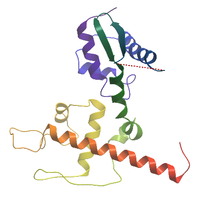
Data Collection: Diffraction data for this dataset was collected from a single crystal at the Diamond synchrotron beamline I04, at a wavelength of 0.9795Å. The data was phased and an initial model was built using the BALBES server. Iterative rounds of manual building using COOT followed by refinement in using autoBUSTER was performed. The deposited structure was refined to a final resolution of 1.62A, R=0.194, Rfree=0.226, and assigned the PDB code 4B4C.
|