KLHL3
PDB:4CH9
Entry Clone Accession:NM_017415.2
Entry Clone Source:MGC
SGC Clone Accession:KLHL3A-c003
Tag:MGHHHHHHSSGVDLGTENLYFQ. N-terminal hexahistidine tag cleavable by TEV protease.
Host:BL21 (DE3)-R3-pRARE2. Phage-resistant derivative of BL21 (DE3), with pRARE2 plasmid encoding rare codon tRNAs (chloramphenicol-resistant).
Vector:pNIC28-Bsa4. T7/lac regulated, N-terminal His-tag, TEV, LIC cloning using BsaI cleavage/T4 polymerase, SacB stuffer fragment, pET28 backbone.
Sequence:
MHHHHHHSSGVDLGTENLYFQSMSLPKVMIVVGGQAPKAIRSVECYDFEEDRWDQIAELPSRRCRAGVVFMAGHVYAVGGFNGSLRVRTVDVYDGVKDQWTSIASMQERRSTLGAAVLNDLLYAVGGFDGSTGLASVEAYSYKTNEWFFVAPMNTRRSSVGVGVVEGKLYAVGGYDGASRQCLSTVEQYNPATNEWIYVADMSTRRSGAGVGVLSGQLYATGGHDGPLVRKSVEVYDPGTNTWKQVADMNMCRRNAGVCAVNGLLYVVGGDDGSCNLASVEYYNPVTDKWTLLPTNMSTGRSYAGVAVIHKSL
Growth
Medium: A glycerol stock was used to inoculate a 10ml starter culture containing LB media with 50µg/ml Kanamycin. The starter culture was grown overnight at 37°C with shaking at 200 rpm. The following morning, flasks containing 1L TB/Kanamycin were each inoculated with 3 ml of the starter culture. Cultures were incubated at 37°C with shaking at 170 rpm until an OD600nm ≥ 2.0 was reached. The flasks were then cooled down to 18°C and 0.4mM IPTG added to induce protein expression overnight. Cells were harvested by centrifugation at 5000 rpm at 4°C for 15 min. Cell pellets from each flask were resuspended in 15ml Binding buffer (50mM HEPES, pH 7.5; 500mM NaCl; 5% Glycerol; 5mM Imidazole) and frozen at -20°C.
Extraction
Procedure:
Extraction buffer, extraction method: The frozen cells were thawed. The cells were lysed by ultrasonication over 15 min with the sonicator pulsing ON for 5 sec and OFF for 10. A final concentration of 0.15% PEI was added to the lysate. The cell lysate was spun down by centrifugation at 21.5K rpm at 4°C for 1 h. The supernatant was recovered for purification. Column 1: Ni-Affinity Chromatography. 5 ml of 50 % Ni-sepharose slurry was applied onto a 1.5 x 10 cm column. The column was equilibrated with binding buffer (50ml). Buffers: Binding buffer: 50 mM Hepes, pH 7.5; 500 mM NaCl; 5% Glycerol; 5 mM imidazole 0.5mM TCEPWash buffer: 50 mM Hepes, pH 7.5; 500 mM NaCl; 5% Glycerol; 25 mM imidazole 0.5mM TCEPElution buffer: 50 mM HEPES, pH 7.5; 500 mM NaCl; 5% Glycerol; 50, 100, 150 and 250 mM imidazole 0.5mM TCEPProcedure: The supernatant following centrifugation was applied by gravity flow onto the Ni-sepharose column. The bound protein was then washed with 50ml binding buffer and subsequently with 30 ml wash buffer. KLHL3A protein was then eluted by applying a step gradient of imidazole - using 5 ml portions of elution buffer with increasing concentration of imidazole (1 x 50 mM, 1 x 100 mM, 1 x 150 mM and 2 x 250 mM). Fractions were analyzed by SDS PAGE and the second, third and fourth elution fractions were kept and pooled. Enzymatic treatment: TEV protease cleavage. Fractions containing KLHL3A were treated with TEV protease overnight at 4°C.Column 2: Anion Exchange Chromatography. HiTrap Q HP 5ml column on ÄKTA-ExpressBuffer: QA Buffer I: 50mM HEPES pH 7.5, 0.5 mM TCEPQB Buffer II: 1M NaCl, 50 mM HEPES pH 7.5, 0.5 mM TCEPProcedure: The HiTrap SP HP column was first washed with 10mL QB buffer II and then equilibrated with 50mL QA buffer I. The protein fraction from above step was concentrated to 5mL using a centrifugal filter with a 10kDa cut-off. The 5mL fraction was made up to 50ml with QA buffer I and applied onto the column. Bound protein was eluted in 0%-100% gradient with QB buffer II. Clean fractions containing the protein were pooled together.Column 3: Size Exclusion Chromatography - S200 HiLoad 16/60 Superdex run on ÄKTA-ExpressGel Filtration Buffer: 300 mM NaCl, 50 mM HEPES pH 7.5, 0.5 mM TCEP, pH 7.5Procedure: The Superdex S200 column was first equilibrated with Gel Filtration buffer. The protein fraction from above step was concentrated to
Structure Determination
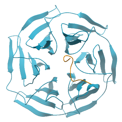
Crystallization:Protein was concentrated to 9 mg/mL. The 11-residue WNK4 peptide (EPEEPEADQHQ) was added to a final concentration of 2 mM. Prior to setting up crystallization plates the solution of KLHL3-WNK4 was incubated on ice for approximately 30 minutes. Sitting-drop vapour diffusion plates were prepared. Crystals grew under multiple conditions using either freshly prepared or frozen protein. The best-diffracting crystals of the KLHL3 complex were obtained by mixing 100 nL of protein with 50 nL of a reservoir solution containing 0.1 M acetate pH 4.3, 0.2 M ammonium sulphate and 25-35 % PEG 4000.. After 3 hours of incubation the drops were spiked with 20 nL of seed-stock solution. Seed stock was prepared from poorly-formed crystals of KLHL3 grown during previous rounds of crystal optimisation, which were diluted in 50-100 µL reservoir solution and vortexed for 2 min in an Eppendorf containing a seed bead. Prior to mounting crystals were cryo-protected in situ by addition of reservoir solution containing an additional 25% ethylene glycol. Crystals were then flash frozen in liquid nitrogen.
Data Collection:1.84 Å resolution; X-ray source: Diamond Light Source, station I02, using monochromatic radiation at wavelength 0.9795 ÅCrystals belonged to spacegroup P212121 with unit cell parameters a=45.33 Å b=84.76 Å c=146.79 Å, α=90° β= 90 γ= 90°. Diffraction data were integrated and scaled using Mofslm and SCALA, respectively. Molecular replacement was used in Phaser MR and PDB entry 2XN4 was used as a search model. Two molecules were present in the asymmetric unit. COOT, REFMAC and PHENIX.REFINE were used for model building and refinement.