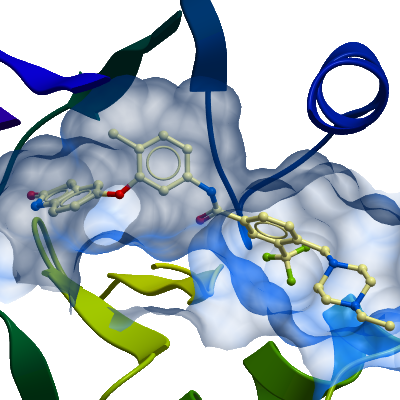
DDR1
PDB:4CKR
Revision
Revision Type:created
Revised by:created
Revision Date:created
Entry Clone Accession:GI:33870807
Entry Clone Source:Mammalian Gene Collection
SGC Clone Accession:DDR1A-c002
Tag:MGHHHHHHSSGVDLGTENLYFQ*S, TEV-cleavable (*) C-terminal hexahistidine tag.
Host:SF9 Spodoptera frugiperda Insect cells
Construct
Prelude:
Sequence:MGHHHHHHSSGVDLGTENLYFQSMPRVDFPRSRLRFKEKLGEGQFGEVHLCEVDSPQDLVSLDFPLNVRKGHPLLVAVKILRPDATKNARNDFLKEVKIMSRLKDPNIIRLLGVCVQDDPLCMITDYMENGDLNQFLSAHQLEDKAAEGAPGDGQAAQGPTISYPMLLHVAAQIASGMRYLATLNFVHRDLATRNCLVGENFTIKIADFGMSRNLYAGDYYRVQGRAVLPIRWMAWECILMGKFTTASDVWAFGVTLWEVLMLCRAQPFGQLTDEQVIENAGEFFRDQGRQVYLSRPPACPQGLYELMLRCWSRESEQRPPFSQLHRFLAEDALNTV
Vector:pFB-LIC-Bse
Growth
Medium:2 L of SF9 cells at a density of 2million/ml were infected with 10ml of Virus/L.Cells were incubated at 27°C in the shaker incubator and harvested after 48 hours. Cells were harvested by centrifugation at 900xG at 4°C for 15 min. Cell pellets from each flask (1l volume) were resuspended in 15ml binding buffer, transferred to 50ml tubes, and stored at -20°C
Antibiotics:
Procedure:
Purification
Buffers
Procedure
Extraction
Buffers
Binding buffer: 50 mM HEPES, pH 7.5; 500 mM NaCl; 5 mM Imidazole; 5% Glycerol.
Procedure
The frozen cells were thawed and protease inhibitor SET V (Calbiochem) added to the cell suspension at 1:1000 dilution. The cells were lysed by high pressure homogenization in an Emulsiflex C5 cell homogeniser. Polyethyleneimin (PEI) was added to a final concentration of 0.5% to precipitate DNA. The cell lysate was clarified by centrifugation at 21,000 rpm at 4°C for 1 h and filtered using syringe filters with a 1.2μm pore size.Column 1: Ni-Affinity Chromatography  2ml Ni-sepharose slurry applied to a 1.5 x 10 cm column.Buffers: Binding buffer: 50mM HEPES, pH 7.5; 500mM NaCl; 5% Glycerol; 5mM Imidazole Wash buffer: 50mM HEPES, pH 7.5; 500mM NaCl; 5% Glycerol; 30mM Imidazole Elution buffer: 50mM HEPES, pH 7.5; 500mM NaCl; 5% Glycerol; 50 to 500mM ImidazoleProcedure: 2ml of 50 % Ni-sepharose slurry (Amersham) was equilibrated in binding buffer and added to the filtered lysate, which was incubated with the Ni-sepharose for 1 hour at 4°C with slow rotation to maximize binding. The lysate was then applied to a 1.5 x 10 cm column by gravity flow. The remaining resin was then washed with 2x50ml binding buffer to remove nonspecifically binding proteins. The bound target protein was eluted by applying a step gradient of imidazole (5 ml fractions of elution buffer supplemented with 50mM and 500mM imidazole). The protein content of collected fractions was visualized using SDS-PAGE and fractions containing DDR1 were pooled. 2 mM DTT and 50 mM L-arginine/50 mM L-glutamate mix were added to the protein for overnight storage.Enzymatic treatment: TEV protease cleavage. Pooled fractions were treated with TEV protease overnight at 4°C.Column 2: Size Exclusion Chromatography  S75 HiLoad 26/60 Superdex run on ÄKTAprime.Buffer: Gel Filtration buffer: 10 mM HEPES pH7.5, 250mM NaCl, 5% Glycerol, 1mM TCEPProcedure: Prior to applying the protein, the S75 26/60 column was washed and equilibrated with gel filtration buffer. Eluted protein from Ni-sepharose column was concentrated to 2ml using an Amicon Ultra-15 filter with a 10kDa cut-off. The concentrated protein was directly applied onto the equilibrated S75 26/60 column, and run at a flow-rate of 2.5 ml/min. 3ml fractions were collected and visualized using SDS-PAGE. Those containing DDR1A were pooled. The final buffer was adjusted to 10 mM HEPES pH 7.5, 250 mM NaCl, 5% glycerol, 1 mM TCEP, 2 mM DTT, 5 mM L-arginine, 5 mM L-glutamate.
Concentration:
Ligand
MassSpec:
Crystallization:DDR1 was co-crystallized with inhibitor at 20°C in 150 nL sitting drops mixing 50 nL protein solution at 8.5 mg/mL with 100 nL of a reservoir solution containing 0.1 M bis-tris propane pH 7.2, 21%(w/v) PEG 3350 and 0.1 M sodium/potassium phosphate. On mounting crystals were cryoprotected with an additional 25% ethylene glycol.
NMR Spectroscopy:
Data Collection:Resolution: 2.2 Ã
X-ray source: Diamond I02Crystals of DDR1A diffracted to a resolution of 2.2 Ã
(scaled resolution). A full dataset was collected at 100 K on Diamond Light Source beamline I02. Crystals belonged to the tetragonal space group P41212. One DDR1 molecule was present in the asymmetric unit. Data were indexed and integrated using XDS and scaled using AIMLESS. Phases were found using molecular replacement in PHASER. PDB entry 3ZOS for use as a search model. The structure was refined and modified using alternate rounds of REFMAC5 and COOT. The final model was validated using MOLPROBITY.
Data Processing: