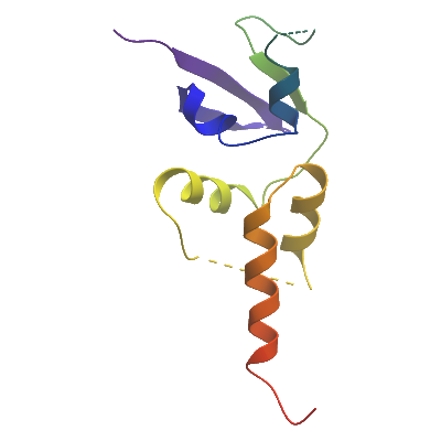
SHKBP1
PDB:4CRH
Revision
Revision Type:created
Revised by:created
Revision Date:created
Entry Clone Accession:BC022855
Entry Clone Source:4CRH
SGC Clone Accession:BC022855
Tag:MGHHHHHHSSGVDLGTENLYFQ. N-terminal hexahistidine tag cleavable by TEV protease.
Host:BL21 (DE3)-R3-pRARE2. Phage-resistant derivative of BL21 (DE3), with pRARE2 plasmid encoding rare codon tRNAs (chloramphenicol-resistant).
Construct
Prelude:
Sequence:MHHHHHHSSGVDLGTENLYFQSMGEVIHLNVGGKRFSTSRQTLTWIPDSFFSSLLSGRISTLKDETGAIFIDRDPTVFAPILNFLRTKELDPRGVHGSSLLHEAQFYGLTPLVRRLQLREELDRSS
Vector:pNIC28-Bsa4. T7/lac regulated, N-terminal His-tag, TEV, LIC cloning using BsaI cleavage/T4 polymerase, SacB stuffer fragment, pET28 backbone.
Growth
Medium:Starter cultures were grown in 10 ml LB + 50 µg/ml kanamycin at 37°C overnight, after inoculation from glycerol stocks. The following day, 7 ml of each starter culture was diluted into 1L LB + 50 µg/ml kanamycin. The 1L culture was grown at 37°C until OD600 reached 0.4-0.5, then the temperature was dropped to 18°C and expression was induced at OD = 0.6-0.8 with 0.4 mM IPTG. Cells were grown at 18°C overnight.The culture was harvested the following day (final OD 4.6) and spun down at 5,000 g and 4°C for 20 min. 15 ml binding buffer (500 mM NaCl, 50 mM HEPES pH 7.5, 5% glycerol, 5 mM imidazole) was added to the cell pellet before storage at -20°C in a 50 ml Falcon tube.
Antibiotics:
Procedure:
Purification
Buffers
Procedure
Extraction
Buffers
Procedure
Cells were thawed and binding buffer was added to each pellet up to 40 ml. Pellets were resuspended by inverting the tubes several times, and 10 μl protease inhibitor mix was added per sample. Cells were sonicated on ice (35% power setting), using a 5 min program (5 s on, 10 s off) (total time 15 min per sample). PEI was added to 0.15% (from a 5% stock solution) to precipitate the DNA. Sonicated cell debris was centrifuged in a JA-25,50 rotor for 45 min at 21,500 rpm and 4°C. The supernatant was cloudy and needed filtering (used a 1.2 μm filter and a syringe).
Column 1: Ni-Affinity Chromatography. 2 ml of 50 % Ni-sepharose slurry was applied onto a 1.5 x 10 cm column. The column was equilibrated with binding buffer (50ml).
Buffers:
Binding buffer: 50 mM Hepes, pH 7.5; 500 mM NaCl; 5% Glycerol; 5 mM imidazoleWash buffer: 50 mM Hepes, pH 7.5; 500 mM NaCl; 5% Glycerol; 25 mM imidazoleElution buffer: 50 mM HEPES, pH 7.5; 500 mM NaCl; 5% Glycerol; 50, 100, 150 and 250 mM imidazole
Procedure: The supernatant following centrifugation was applied by gravity flow onto the Ni-sepharose column. The bound protein was then washed with 50 ml binding buffer and subsequently with 30 ml wash buffer. SHKBP1 protein was then eluted by applying a step gradient of imidazole - using 5 ml portions of elution buffer with increasing concentration of imidazole (1 x 50 mM, 1 x 100 mM, 1 x 150 mM and 2 x 250 mM). Fractions were analyzed by SDS PAGE and the second, third and fourth elution fractions were kept and pooled.
Enzymatic treatment: TEV protease cleavage. Fractions containing SHKBP1 were treated with TEV protease overnight at 4°C.
Column 2: Size Exclusion Chromatography - S200 HiLoad 16/60 Superdex run on ÄKTA-Express
Gel Filtration Buffer: 300 mM NaCl, 50 mM HEPES pH 7.5, 0.5 mM TCEP, 5% glycerol
Procedure: The Superdex S200 column was first equilibrated with Gel Filtration buffer. The protein fraction from above step was concentrated to Concentration:
Ligand
MassSpec:
Crystallization:The protein was buffer exchanged gradually during concentration (NaCl, DTT and Arg-Glu were added to avoid precipitation), to a final buffer composition of: 400 mM NaCl, 50 mM HEPES pH 7.5, 0.5 mM TCEP, 2.5% glycerol, 5 mM DTT, 20 mM Arg-Glu. The protein was concentrated to 7.7 mg/ml. Sitting-drop vapour diffusion plates were prepared. Crystals grew under multiple conditions using either freshly prepared or frozen protein. Several crystals were mounted. The best-diffracting crystals of SHKBP1 were obtained by mixing 50 nl protein with 100 nl of a reservoir solution containing 17% PEGm2K, 0.1 M Tris pH 8.4, 0.15 M TMAO at 20⁰C.
NMR Spectroscopy:
Data Collection:1.72 Å resolution; X-ray source: Diamond Light Source, station I04, using monochromatic radiation at wavelength 0.9795 Å
Data Processing: