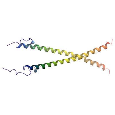
ERCC6
PDB:4CVO
Revision
Revision Type:created
Revised by:created
Revision Date:created
Entry Clone Accession:NM_000124
Entry Clone Source:SGC Clone Accession:ERCC6A-c055
Tag:Tag TEV-cleavable N-terminal his6 tag
Host:BL21(DE3)-R3-pRARE2
Construct
Prelude:Sequence:mhhhhhhssgvdlgtenlyfq^SMEPSAQALELQGLGVDVYDQDVLEQGVLQQVDNAIHEASRASQLVDVEKEYRSVLDDLTSCTTSLRQINKIIEQLSPQ
Vector:pNIC28-Bsa4. Details [
PDF ]; Sequence [
FASTA ] or [
GenBank ].
Growth
Medium:Antibiotics:Procedure:Growth medium, induction protocol:The expression plasmid was transformed into the host strain and plated on LB-agar containing 50 µg/ml kanamycin and 34 µg/ml chloramphenicol. Several colonies were combined to inoculate a 1ml culture in TB (+ 50 µg/ml kanamycin, 34 µg/ml chloramphenicol). The culture was grown overnight, glycerol was added to 15% v/v (from a 50% stock), and the resulting glycerol stock was frozen at -80°C in 100 µl aliquots. A loopful of cells from the glycerol stock was inoculated into 10-ml of TB medium containing 50 µg/ml kanamycin and 34 µg/ml chloramphenicol and grown overnight at 37°C. 800 ml of TB medium (containing 50 µg/ml kanamycin) was inoculated with 10 ml of the overnight culture and grown in 2 L straight walled flaskes until OD600 of 2.0. Cells were cooled to 18°C, IPTG added to 1 mM and growth continued at 18°C overnight. The cells were collected by centrifugation then the pellets were scraped out and transferred to 50-ml Falcon tubes and frozen at -80°C.
Purification
BuffersProcedureExtraction
BuffersProcedureCell extraction : Frozen cell pellets (approx. 6g) were thawed briefly in a bath of warm water (20 - 37°C) then transferred to ice. Six volumes (i.e. 6 ml for every gram of cells) of lysis buffer was added, and the cells were resuspended by agitating and disrupted by pulsed sonication (5 sec on 10 sec off) for 15 minutes on ice. lysates were centrifuged for 25 minutes at 65,000xg to remove cellular debris.
Lysis buffer: 50 mM HEPES, pH 7.5, 500 mM NaCl, 5% glycerol, 20 mM Imidazole.
Column 1 : 2ml bed volume Ni-IDA
Solutions: Wash buffer: 50 mM HEPES, pH 7.5, 500 mM NaCl, 45 mM imidazole, 5% Glycerol.
Elution buffer: 50 mM HEPES, pH 7.5, 500 mM NaCl, 300 mM imidazole, 5% Glycerol.
Procedure: The cell extract was incubated with 5ml of Ni-IDA resin (GE Healthcare) for 1hr at 4°c with gentle agitation. The resin was pelleted by centrifugation (700g for 6 min), and the supernatant was decanted. The resin was washed initially with 10 column volumes of lysis buffer and pelleted by centrifugation. Ten column volumes of wash buffer1 were added and the resuspended resin was transferred to a gravity flow column and washed with 5 column volumes of wash buffer. Proteins were eluted by addition of 2 x 10 ml of elution buffer and combined with the fractions from the wash buffer.Enzymatic treatment and Column 2: The N-terminal His6-tag was cleaved by incubating the protein overnight with tobacco etch virus (TEV) protease (1:40 ratio at 4°C) whilst being dialyzed into dialysis buffer (50 mM HEPES, 500 mM NaCl, 5 % Glycerol,) using a 3,500 MWCO snakeskin dialysis membrane. Cleaved protein was purified by passing over a 5 ml pre-equilibriated 50% Ni-IDA bead solution. Elution was done in GF buffer supplemented with step gradients of 20 mM, 40 mM 100 mM and 300 mM imidazole, with flowthrough and fractions collected.
Column 3 : Gel filtration, Hiload 16/60 Superdex S75 prep grade, 120 ml (GE Healthcare)
GF buffer: 50 mM Tris, pH 8, 250 mM NaCl.
Procedure: The fractions from Ni-IDA column were concentrated to 2ml using a 3 KDa MWCO centrifugal concentrator and loaded on the gel filtration column in GF buffer at 1 ml/min. Eluted proteins were collected in 2-ml fractions and analysed on SDS-PAGE. ERCC6A eluted as two distinct peaks from the gel filtration column consistent with dimeric and tetrameric species. Both peaks were used in crystallization experiments separately, with the tetrameric peak (eluting at approx. 58ml) producing the final crystals used for structure determination.
Concentration:Concentration method: The cleaved purified protein was concentrated using a 3 KDa MWCO centrifugal concentrator to 13 mg/ml and stored at 4°C. The protein concentration was determined spectrophotometrically using ε280 = 2980
LigandMassSpec:Observed mass of protein was 17515.0, theoretical mass is 8757.7, thus the mass is correct for a disulphide bonded dimer.
Crystallization:Crystallisation experiments were performed using the sitting drop vapour diffusion technique. Crystals of ERCC6A grew at 20°C from conditions containing 1.6M Magnesium Sulfate -- 0.1M MES pH 6.5, and were cryo protected by transferring to a cryo solution consisting of the well solution supplemented with the addition of 25% Ethelyene glycol before being plunged into liquid nitrogen.
NMR Spectroscopy:Data Collection:Data was collected at diamond light source beamline I03. Data were processed using XDS, and the structure was solved by molecular replacement using an idealized alpha helical model and the program PHASER. Model building was performed with the program COOT and the model refinement was performed using REFMAC to a final Rfactor=23.7 % , Rfree =25.5 %.
Data Processing: