BAZ2A
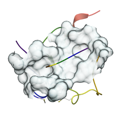
PDB:4Q6F
Revision
Revision Type:created
Revised by:created
Revision Date:created
Entry Clone Accession:BAZ2A_HUMAN Q9UIF9
Entry Clone Source:synthetic
SGC Clone Accession:
Tag:MCSSHHHHHHGSGSGSDQEAKPSTEDLGDKKEGEYIKLKVIGQDSSEIHFKVKMTTHLKKLKESYCQRQGVPMNSLRFLFEGQRIADNHTPKELGMEEEDVIEVYQEQTGG(*)H (*) N-terminal his6 tag + SUMO tag
Host:BL21(DE3)-R3-pRARE2
Construct
Prelude:
Sequence:MSVNKVTCLVCRKGDNDEFLLLCDGCDRGCHIYCHRPKMEAVPEGDWFCTVCLAQQV
Vector:pET15
Growth
Medium:
Antibiotics:
Procedure:Best expression construct BAZ2A-c026 (AmpR) was transformed into Escherichia coli competent BL21(DE3) Rosetta (ChlR) cells or into BL21 (DE3)-R3-pRARE2 (ChlR) cells (phage-resistant derivative with a pRARE plasmid encoding rare codon tRNAs). Cells were grown at 37°C in Terrific Broth from overnight cultures until A600 reached between 0.6-0.8, then the media was cooled and 0.2 mM isopropyl-β-Dthiogalactopyranoside (IPTG) and an extra 0.1 mM ZnCl2 were added to induce the protein expression at 20°C for 16 hours
Purification
Buffers
Procedure
STEP1: after centrifugation, the supernatant was loaded onto the nickel column and eluted in an imidazole linear gradient. Binding buffer 20 mM HEPES pH 7.5; 500 mM NaCl; 10 mM imidazole, 0.5 mM TCEP; Elution buffer 20 mM HEPES pH 7.5; 500 mM NaCl; 500 mM imidazole and 0.5 mM TCEP.STEP2: the eluted protein was collected and treated overnight with SENP1 (sumo endoprotease 1) protease at 4ºC to remove the N terminal tag. Dialysis buffer 20 mM Hepes pH 7.5, 500 mM NaCl, 0.5 mM TCEP (no imidazole)STEP3: digested protein was loaded onto a nickel column again to remove the cleaved 6His-SUMO tag and the hexa-histidine expression SENP1 protease used. The flow through containing the untagged protein was collected and further dialysed. Binding buffer 20 mM Hepes pH 7.5, 500 mM NaCl, 0.5 mM TCEP, 20 mM imidazole; Elution buffer 20 mM Hepes pH 7.5, 500 mM NaCl, 0.5 mM TCEP, 500 mM ImidazoleSTEP4: a size exclusion chromatography (HiLoad 16/600 Superdex 75) is performed to remove soluble aggregates, buffer 20 mM Hepes pH 7.5, 200 mM NaCl, 2 mM DTT.STEP5: the last polishing step is through ion exchange chromatography (RESOURCE Q 6 ml). Protein was eluted in a linear gradient of NaCl. Binding buffer 20 mM Hepes pH 7.5, 5 mM NaCl, 2 mM DTT. Elution buffer 20 mM Hepes pH 7.5, 500 mM NaCl, 2 mM DTT.
Extraction
Buffers
Procedure
Frozen pellet was thawed and resuspended in binding buffer containing 20 mM HEPES pH 7.5; 500 mM NaCl; 10 mM imidazole and 0.5 mM TCEP. Cells were lysed using an EmulsiFlex-C5 high-pressure homogenizer (Avestin - Mannheim, Germany) or high-pressure cell disruption (Constant Systems Limited) in binding buffer from the nickel affinity column (HisTrap Chelating FF 5ml) in the presence of Protease Inhibitor Cocktail EDTA-free. Lysates were cleared by centrifugation (14,000 x g for 45h at 4ºC, JLA 16.250 rotor, on a Beckman Coulter Avanti J-20 XP centrifuge).
Concentration:The cleaved purified protein was concentrated in a VivaSpin (3 K MWCO) to 6.7 mg/ml prior to crystallization trials. The protein concentration was determined spectrophotometrically.
Ligand
MassSpec:
Crystallization:Histone peptide H3K4 complex crystallization with BAZ2A PHD finger: peptide was soaked into pre-formed apo BAZ2A PHD finger crystals grown in 2 M malic acid pH 7 as crystallization condition. Soaking solution contained 1.6 M malic acid and 12 mM H3K4 5-mer peptide and the crystals were soaked for > 12h at 20ºC. Crystals were cryo-protected in 1.4 M malic acid, 6 mM H3K4 and 20% ethyleneglycol
NMR Spectroscopy:
Data Collection:A dataset was collected for the soaked crystal with H3K4 histone peptide at Diamond Light Source (beamline I03) and processed to 1.91 Å using XDS and AIMLESS. The crystal structure was solved by molecular replacement using PHASER with the apo BAZ2A PHD finger pdb accession code 4QF2.pdb as the searching templates. The coordinate solution was further manually built and refined using Coot and REFMAC5.
Data Processing: