RPRD1B
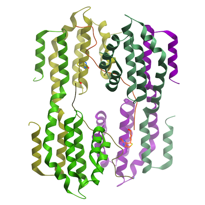
PDB:4Q96
Revision
Revision Type:created
Revised by:created
Revision Date:created
Entry Clone Accession:NM_021215.3
Entry Clone Source:MGC FL RPRD1B (NM_021215.3)
SGC Clone Accession:DCC020:G07
Tag:N-terminal tag: MHHHHHHSSGRENLYFQG
Host:BL21 (DE3) Codon plus RIL (Stratagene)
Construct
Prelude:Sequence:MHHHHHHSSGRENLYFQGGSSFSESALEKKLSELSNSQHSVQTLSLWLIHHRKHAGPIVSVWHRELRKAKSNRKLTFLYLANDVIQNSKRKGPEFTREFESVLVDAFSHVAREADEGCKKPLERLLNIWQERSVYGGEFIQQLKLSMEDSKSP
Vector:pET28-MHL
Growth
Medium:Antibiotics:Procedure:A 250 mL flask containing LB (Sigma L7658) supplemented with 50 µg/mL kanamycin (BioShop Canada KAN 201) was inoculated from a glycerol stock of the bacteria. The flask was shaken overnight (16 hours) at 250 rpm at 37 ºC. Using the Lex system, a 2L bottle (VWR 89000-242) containing 1800 mL of TB (Sigma T0918) supplemented with 1.5% glycerol, 50 ug/ mL kanamycin and 600 µl antifoam 204 (Sigma A-8311) was inoculated with 50 mL overnight LB culture, and incubated at 37 ºC. The temperature of the media was reduced to 15 ºC one hour prior to induction and induced at OD600 = 6 with 100 µM isopropyl-thio-β-D-galactopyranoside (BioShop Canada IPT 001). Cultures were aerated overnight (16 hours) at 18 ºC, and cell pellets collected by centrifugation and frozen at -80 ºC.
Purification
ProcedureIMAC: Unclarified lysate was mixed with 2-3 mL of Ni-NTA superflow Resin (Qiagen) per 40 mL lysate. The mixture was incubated with mixing for at least 45 minutes at 4oC. The mixture was then loaded onto an empty comLum (BioRad) and washed with 100 mL wash buffer. Samples were eluted from the resin by exposure to 2-3 column volumes (approx. 10-15 mL) of elution buffer. Concentration of eluted protein was estimated by OD280. pTEV was added to eluted protein at 1:20 for eluted protein and dialyze against gel filtration buffer overnight to remove His-tag.
Gel filtration chromatography: An XK 26x65 column (GE Healthcare) packed with HighLoad Superdex 75 resin (GE Healthcare) was pre-equilibrated with gel filtration buffer for 1.5 column volumes using an AKTA explorer (GE Healthcare) at a flow rate of 1.0 mL/min. The dialyzed sample from the IMAC step (approx. 15 mL) was loaded onto the column at 1.5 mL/min, and 2mL fractions were collected into 96-well plates (VWR 40002-012) using peak fractionation protocols). Fractions observed by a UV absorption chromatogram to contain the protein were pooled.
Extraction
ProcedureFrozen cell pellet contained in bags (Beckman 369256) obtained from 2L of culture were thawed by soaking in warm water. Each cell pellet was resuspended in 25-40 mL lysis buffer and homogenized using an Ultra-Turrax T8 homogenizer (IKA Works) at maximal setting for 30-60 seconds per pellet. Cell lysis was accomplished by sonication (Virtis408912, Virsonic) on ice: the sonication protocol was 10 sec pulse at half-maximal frequency (5.0), 10 second rest, for 10 minutes total sonication time per pellet.
Concentration:Purified proteins were concentrated using 15 mL concentrators with a 10,000 molecular weight cut-off (Amicon Ultra-15, UFC900524, Millipore) at 3750 rpm, typically resulting in a final concentration around 20 mg/mL.
LigandMassSpec:Crystallization:Crystal complex of RPRD1B CID with Ser2P CTD peptide (1:3) was grown in 2.0 M ammonium sulfate, 5% isopropanol, vapor diffusion, at temperature 291K; Crystal complex of RPRD1B CID with unmodified CTD peptide (1:5) was grown in 25% PEG-1500, 0.2M ammonium sulfate, 0.1M HEPES, pH 7.5, vapor diffusion at temperature 291K.
Crystals grow to a mountable size in several days.Cryo contains mother liquid with 10% Glycerol.
NMR Spectroscopy:
Data Collection:
Data Processing: