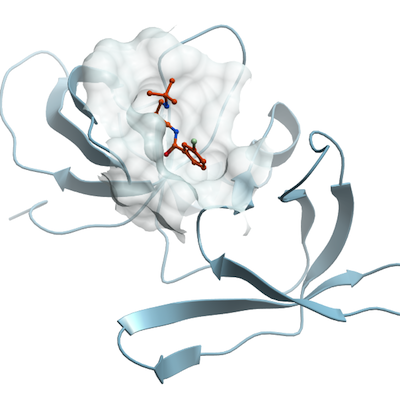
TP53BP1
PDB:4RG2
Revision
Revision Type:created
Revised by:created
Revision Date:created
Entry Clone Accession:
Entry Clone Source:
SGC Clone Accession:APC056:E04
Tag:N-terminal His tag folowed by a TEV cleavage site
Host:BL21(DE3)-V2R-pRARE2
Construct
Prelude:
Sequence:MHHHHHHSSGRENLYFQGGNSFVGLRVVAKWSSNGYFYSGKITRDVGAGKYKLLFDDGYECDVLGKDILLCDPIPLDTEVTALSEDEYFSAGVVKGHRKESGELYYSIEKEGQRKWYKRMAVILSLEQGNRLREQYGLGPYE
Vector:pET28-MHL
Growth
Medium:
Antibiotics:
Procedure:
Purification
Procedure
The cell pellet (18.8 g) was thawed and resuspended in 190 mL of lysis buffer (20 mM HEPES, pH 7.5, 500 mM NaCl, 5 mM imidazole, 0.5 mM TCEP, and 5% glycerol). The cell suspension was supplemented with 0.5% (w/v) CHAPS, 5 ul of benzonase (EMD Millipore, cat. no. 70746), protease inhibitor cocktail (Roche) and the cells were sonicated on ice for 5 min total (10 s pulses with 5s interruptions). The lysate was clarified by centrifugation at 20,000 × g, 4 °C, 60 min and the resulting supernatant was filtered through 0.45 um filter and applied onto 5 mL HisTrap HP column (GE). The column was washed with 10 CV of wash buffer (20 mM HEPES, pH 7.5, 500 mM NaCl, 40 mM imidazole, 0.5 mM TCEP, and 5% glycerol) and the protein was eluted using elution buffer (20 mM HEPES, pH 7.5, 500 mM NaCl, 250 mM imidazole, 0.5 mM TCEP, and 5% glycerol). TEV protease was added to the eluted protein (at ratio of 1 mg of TEV protease per 50 mg protein), and incubated overnight at 4 °C during dialysis against 20 mM HEPES, pH 7.5, 500 mM NaCl, 0.5 mM TCEP, and 5% glycerol. The uncleaved protein (and His-tagged TEV protease) were removed by passing through 1 mL HisTrap HP column (GE). The cleaved protein was collected in the flow-through fraction. As the final purification step, the cleaved protein was applied on 16/600 Superdex 200 (GE) column equilibrated with 20 mM HEPES, pH 7.5, 150 mM NaCl, 0.5 mM TCEP. Final purification yield was 25 mg of protein per 1 L of culture and the purity of the protein was over 95%. The MW (14,103.9 Da) of the purified construct was confirmed by LC/MSD TOF (Agilent).
Extraction
Procedure
The expression construct for 53BP1 tudor domain (residues 1438 - 1606) in pET28-MHL vector was transformed into BL21(DE3)-V2R-pRARE2 cells. The cells were incubated in Terrific Broth medium (TB) in the presence of 50 ug/mL kanamycin and 34 ug/mL chloramphenicol at 37 °C. When the OD600 reached 1.5, the overexpression of 53BP1 was induced by addition of isopropyl-1-thio-D-galactopyranoside (IPTG), final concentration 0.5 mM, and incubated overnight at 16 °C. Next day, the cells were harvested by centrifugation at 12,227 × g (10 min, 4C) and the cell pellets were flash frozen in liquid N2 and stored at -80 °C.
Concentration:
Ligand
MassSpec:
Crystallization:Purified 53BP1 (42 mg/mL) in 20 mM HEPES, pH 7.5, 150 mM NaCl, 0.5 mM TCEP was pre-incubated with 10 mM UNC2170 (dissolved in water) and the best crystals were obtained by vapor diffusion technique at 20 °C in sitting drops by mixing 1 uL of protein solution with 1 uL of reservoir solution containing 19% PEG3350, 150 mM DL-malic acid pH 7.2. For cryoprotection, the crystals were soaked in the reservoir solution supplemented with 15% ethylene glycol (v/v) for 60 s before flash freezing in liquid N2.
NMR Spectroscopy:
Data Collection:
Data Processing: