Molecular Biology
Entry Clone Accession: NP_061852
Entry Clone Source: Dundee MRC-PPU
SGC Construct ID: WNK1A-c011
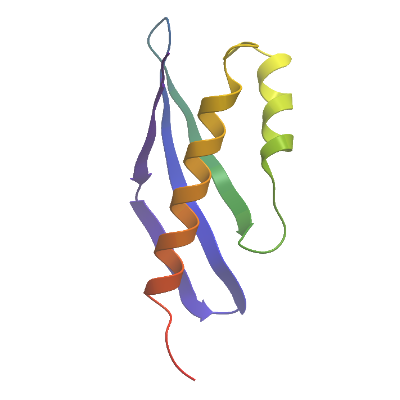
Protein Region: T1115-E1201
Vector: pNIC28-Bsa4
Tag: N-6HIS;N-TEV
Host: BL21(DE3)-R3-pRARE2
Sequence (with tag(s)): MHHHHHHSSGVDLGTENLYFQSMTSRPKLRILNVSNKGDRVVECQLETHNRKMVTFKFDLDGDNPEEIATIMVNNDFILAIERESFVDQVREIIEKADEMLSEDVSVEPE
Sequence after tag cleavage: SMTSRPKLRILNVSNKGDRVVECQLETHNRKMVTFKFDLDGDNPEEIATIMVNNDFILAIERESFVDQVREIIEKADEMLSEDVSVEPE
DNA Sequence: CATATGCACCATCATCATCATCATTCTTCTGGTGTAGATCTGGGTACCGAGAACCTGTACTTCCAATCCATGACTTCACGCCCAAAATTAAGAATTTTGAATGTTTCAAATAAAGGAGACCGAGTAGTAGAATGTCAATTAGAGACTCATAATAGGAAAATGGTTACATTCAAATTTGACCTAGATGGTGACAACCCCGAGGAGATAGCAACAATTATGGTGAACAATGACTTTATTCTAGCAATAGAGAGAGAGTCGTTTGTGGATCAAGTGCGAGAAATTATTGAAAAAGCTGATGAAATGCTCAGTGAGGATGTCAGTGTGGAACCAGAGTGACAGTAAAGGTGGATACGGATCCGAA
Protein Expression
Medium: LB
Antibiotics: Kanamycin and Chloramphenicol
Procedure: A glycerol stock was used to inoculate a 15ml starter culture of LB with 50ug/ml Kanamycin and 34 ug/ml Chloramphenicol. The starter culture was grown overnight at 37⁰C, shaking at 220rpm. The starter culture was used to inoculate 1l cultures supplemented with Kanamycin and Chloramphenicol at 5ml/l which were then grown at 37⁰C, shaking at 180rpm, until reaching an optical density of 0.6. Cultures were induced with 0.4mM Isopropyl β-D-1-thiogalactopyranoside and cooled to 18⁰C and left shaking overnight at 180rpm. Cultures were harvested by centrifugation at 5000g for 15 mins. Cell pellets were resuspended in 50 mM HEPES pH 7.5, 500 mM NaCl, 5 mM Imidazole, 5 % glycerol plus Merck Set III protease inhibitor and stored at -20°C.
Protein Purification
Procedure: Cells were lysed by sonication and lysates clarified by centrifugation at 21,000rpm following the addition of 0.125% polyethyleneimine. The resulting supernatant was incubated with Ni-IMAC resin (equilibrated in 50 mM HEPES pH 7.5, 500 mM NaCl, 5 mM Imidazole, 5 % glycerol), mixing by inversion, at 4⁰C for 1 hour. This was centrifuged at 700g for 5mins, the supernatant removed and resin resuspended in 50 mM HEPES pH 7.5, 500 mM NaCl, 5 mM Imidazole, 5 % glycerol. This was applied to a gravity column and washed and eluted with 50 mM HEPES pH 7.5, 500 mM NaCl, 5% glycerol, and 30-250mM imidazole. Fractions containing protein (as seen by SDS-PAGE) were treated with TEV protease overnight at 4⁰C prior to gel filtration on an S75 gel filtration column using 50 mM HEPES pH 7.5, 300 mM NaCl and 1mM TCEP as the running buffer. Fractions containing purified protein (as observed by SDS-PAGE) were concentrated to 10.0 mg/ml using a centrifugal concentrator. Protein concentration was determined by Bradford Assay due to low extinction coefficient.
Columns: Column 1: Ni drip column; Column 2: S75;
Concentration: 10.0 mg/ml
Mass-spec Verification: Intact mass confirmed by LC-MS as 12735.6 Da.
Structure Determination
Crystallization: Protein was incubated with the peptide RGRFQVITIPQ dissolved in DMSO to a concentration of 1mM, at a ratio of 1:3. Crystals were grown using the sitting drop vapor diffusion method at 20⁰C. Crystals were grown in 150 nl drops consisting of 1:2 mother liquor (20% PEG3350, 10% ethylene glycol, 0.1M bis-tris-propane pH 6.5, 0.2M sodium formate) to protein (7 mg/ml) with peptide. 25% ethylene glycol was added to the crystal as a cryoprotectant and the crystal was mounted and flash frozen in liquid nitrogen. The protein crystallised with the C-terminal peptide in the binding pocket rather than the peptide which was added.
Data Collection: Data was collected at beamline I04-1 at 100K.
Data Processing: Data was processed to a resolution of approximately 1.6 Å using XDS and either xia2 or autoPROC, or DIALS.