No. XX AAK1A PDB: 5L4Q Materials & Methods |
Entry Clone Source: MGC |
Entry Clone Accession: |
SGC Construct ID: AAK1A-c046 |
Coding DNA sequence:
ATGACCTCGGGCCTGGGCAGTGGCTA
CATCGGAAGAGTCTTCGGCATCGGGC
GACAGCAGGTCACAGTGGACGAGGTG
TTGGCGGAAGGTGGATTTGCTATTGT
ATTTCTGGTGAGGACAAGCAATGGGA
TGAAATGTGCCTTGAAACGCATGTTT
GTCAACAATGAGCATGATCTCCAGGT
GTGCAAGAGAGAAATCCAGATAATGA
GGGATCTTTCAGGGCACAAGAATATT
GTGGGTTACATTGATTCTAGTATCAA
CAACGTGAGTAGCGGTGATGTATGGG
AAGTGCTCATTCTGATGGACTTTTGT
AGAGGTGGCCAGGTGGTAAACCTGAT
GAACCAGCGCCTGCAAACAGGCTTTA
CAGAGAATGAAGTGCTCCAGATATTT
TGTGATACCTGTGAAGCTGTTGCCCG
CCTGCATCAGTGCAAAACTCCTATTA
TCCACCGGGACCTGAAGGTTGAAAAC
ATCCTCTTGCATGACCGAGGCCACTA
TGTCCTGTGTGACTTTGGAAGCGCCA
CCAACAAATTCCAGAATCCACAAACT
GAGGGAGTCAATGCAGTAGAAGATGA
GATTAAGAAATACACAACGCTGTCCT
ATCGAGCACCAGAAATGGTCAACCTG
TACAGTGGCAAAATCATCACTACGAA
GGCAGACATTTGGGCTCTTGGATGTT
TGTTGTATAAATTATGCTACTTCACT
TTGCCATTTGGGGAAAGTCAGGTGGC
AATTTGTGATGGAAACTTCACAATTC
CTGATAATTCTCGATATTCTCAAGAC
ATGCACTGCCTAATTAGGTATATGTT
GGAACCAGACCCTGACAAAAGGCCGG
ATATTTACCAGGTGTCCTACTTCTCA
TTTAAGCTACTCAAGAAAGAGTGCCC
AATTCCAAATGTACAGAACTCTCCCA
TTCCTGCAAAGCTTCCTGAACCAGTG
AAAGCCAGTGAGGCAGCTGCAAAAAA
GACCCAGCCAAAGGCCAGACTGACAG
ATCCCATTCCCACCACAGAGACTTCA
ATTGCAGCAGAGAACCTCTACTTCCA
ATCGCACCATCATCACCACCATTGA
|
Expressed protein sequence:
MTSGLGSGYIGRVFGIGRQQVTVDEV
LAEGGFAIVFLVRTSNGMKCALKRMF
VNNEHDLQVCKREIQIMRDLSGHKNI
VGYIDSSINNVSSGDVWEVLILMDFC
RGGQVVNLMNQRLQTGFTENEVLQIF
CDTCEAVARLHQCKTPIIHRDLKVEN
ILLHDRGHYVLCDFGSATNKFQNPQT
EGVNAVEDEIKKYTTLSYRAPEMVNL
YSGKIITTKADIWALGCLLYKLCYFT
LPFGESQVAICDGNFTIPDNSRYSQD
MHCLIRYMLEPDPDKRPDIYQVSYFS
FKLLKKECPIPNVQNSPIPAKLPEPV
KASEAAAKKTQPKARLTDPIPTTETS
IAAENLYFQSHHHHHH
|
Vector: pNIC-CTH0
C-terminal His, TEV-cleavable, for LIC cloning.
|
Tags and additions: AENLYFQSHHHHHH: C-terminal hexahistidine tag cleavable by TEV protease. |
Host: BL21 (DE3)-R3-pRARE2. Phage-resistant derivative of BL21 (DE3), with pRARE2 plasmid encoding rare codon tRNAs (chloramphenicol-resistant). |
Growth Medium & Induction Protocol: A glycerol stock was used to inoculate a 10ml starter culture containing LB media with 50µg/ml Kanamycin + 34 µg/ml Chloramphenicol . The starter culture was grown overnight at 37°C with shaking at 200 rpm. The following morning, flasks containing 1L TB/Kanamycin were each inoculated with 3 ml of the starter culture. Cultures were incubated at 37°C with shaking at 170 rpm until an OD600nm ⥠1.4 was reached. The flasks were then cooled down to 18°C and 0.4mM IPTG added to induce protein expression overnight at OD600nm ⥠2.0. Cells were harvested by centrifugation at 5000 rpm at 4°C for 15 min. Cell pellets from each flask were resuspended in 15ml Binding buffer (50mM HEPES, pH 7.5; 500mM NaCl; 5% Glycerol; 5mM Imidazole; 0.5mM TCEP; 1:2000 Protease Inhibitor Cocktail) and frozen at -20°C. |
Extraction buffer, extraction method: The frozen cells were thawed. The cells were lysed by ultrasonication over 15 min with the sonicator pulsing ON for 5 sec and OFF for 10. A final concentration of 0.15% PEI was added to the lysate. The cell lysate was spun down by centrifugation at 22 K rpm at 4°C for 1 h. The supernatant was recovered for purification.
|
Column 1: Ni-Affinity Chromatography. 5 ml of 50 % Ni-sepharose slurry was applied onto a 1.5 x 10 cm column. The column was equilibrated with binding buffer (50ml). |
Buffers:
Binding buffer: 50 mM Hepes, pH 7.5; 500 mM NaCl; 5% Glycerol; 5 mM imidazole 0.5mM TCEP
Wash buffer: 50 mM Hepes, pH 7.5; 500 mM NaCl; 5% Glycerol; 30 mM imidazole 0.5mM TCEP
Elution buffer: 50 mM HEPES, pH 7.5; 500 mM NaCl; 5% Glycerol; 50, 100, 150 and 250 mM imidazole 0.5mM TCEP
|
Procedure: Carried out in a cold room. The supernatant following centrifugation was applied by gravity flow onto the Ni-sepharose column. The bound protein was then washed with 50ml binding buffer and subsequently with 30 ml wash buffer. AAK1A protein was then eluted by applying a step gradient of imidazole - using 5 ml portions of elution buffer with increasing concentration of imidazole (1 x 50 mM, 1 x 100 mM, 1 x 150 mM and 2 x 250 mM). Fractions were analyzed by SDS PAGE and the first, second, third & fourth elution fractions were kept and pooled. |
Enzymatic treatment: TEV protease cleavage. Fractions containing AAK1A were treated with TEV protease overnight at 4°C. |
Column 2: Size Exclusion Chromatography - S75 HiLoad 16/60 Superdex run on ÄKTA-Express |
Gel Filtration buffer: 300 mM NaCl, 50 mM HEPES pH 7.5, 0.5 mM TCEP, pH 7.5, 5% glycerol
|
Procedure: Column stored in a cold room. The Superdex S75 column was first equilibrated with Gel Filtration buffer. The protein fraction from above step was concentrated to |
Column 3: Ni-Affinity Chromatography. 1 ml of 50 % Ni-sepharose slurry was applied onto a 1.0 x 10 cm column. The column was equilibrated with binding buffer (15ml). |
Buffers:
Binding buffer: 50 mM Hepes, pH 7.5; 500 mM NaCl; 5% Glycerol; 5 mM imidazole 0.5mM TCEP
Wash buffer: 50 mM Hepes, pH 7.5; 500 mM NaCl; 5% Glycerol; 30 mM imidazole 0.5mM TCEP
Elution buffer: 50 mM HEPES, pH 7.5; 500 mM NaCl; 5% Glycerol; 250 mM imidazole, 0.5mM TCEP
|
Procedure: Carried out at room temperature, with fractions stored on ice immediately following collection. The pooled fractions from gel filtration were applied by gravity flow onto the Ni-sepharose column and flow-through collected. The column was then washed with 4ml wash buffer and subsequently with 5ml elution buffer. Fractions were analyzed by SDS PAGE and the flow-through containing AAK1A was used for crystallization. |
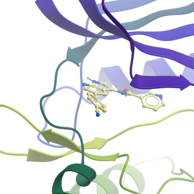
Crystallization: Dephosphorylated protein was concentrated to 12 mg/mL. The compound LKB1 (AAK1 dual inhibitor) was added to a final concentration of 1.5 mM from a stock at 50 mM in 100% DMSO (3% final DMSO concentration). Prior to setting up crystallization plates the solution of AAK1-LKB1 was incubated on ice for approximately 30 minutes, then centrifuged at 14,000 rpm for 10 minutes, 4°C. Sitting-drop vapour diffusion plates were prepared. Crystals grew under multiple conditions using freshly prepared protein. The best-diffracting crystals of the AAK1-LKB1 complex were obtained using a reservoir solution containing 26% PEG 3350, 0.1 M bis-Tris pH5.5 by spiking drops with 20 nL of seed-stock solution immediately prior to incubation at 18°C. Seed stock was prepared from poorly-formed crystals of AAK1-LKB1 grown during previous rounds of crystal optimisation, which were diluted in 50-100 µL reservoir solution and vortexed for 2 min in an Eppendorf containing a seed bead. A 1:1 dilution series of seeds was prepared in order to find the optimal seed concentration. Prior to mounting crystals were cryo-protected in situ by addition of reservoir solution containing an additional 25% ethylene glycol. Crystals were then flash frozen in liquid nitrogen. |
Data Collection:2 Å resolutionX-ray source: Diamond Light Source, station I02, using monochromatic radiation at wavelength 0.9795 Å
Crystals belonged to spacegroup P1211 with unit cell parameters a=61.8 Å b=55.05 Å c=86.57 Å, α=90° β= 104.44 &= 90°. Diffraction data were integrated and scaled using xia2 & Aimless. Molecular replacement was used in Phaser MR and PDB entry 4wsq was used as a search model. Two molecules were present in the asymmetric unit. COOT, REFMAC and PHENIX.REFINE were used for model building and refinement.
|