Molecular Biology
Entry Clone Accession: IMAGE:3048375
Entry Clone Source: MGC
SGC Construct ID: DCLRE1AA-c081
Protein Region: K698-Y1040
Vector: pFB-LIC-Bse. This is a baculovirus transfer vector (Bac-to-bac), with N-terminal 6 His tag followed by a TEV cleavage site
Host: DH10Bac
Sequence (with tag(s)): MGHHHHHHSSGVDLGTENLYFQSMKKTCPFYKKIPGTGFTVDAFQYGVVEGCTAYFLTHFHSDHYAGLSKHFTFPVYCSEITGNLLKNKLHVQEQYIHPLPLDTECIVNGVKVVLLDANHCPGAVMILFYLPNGTVILHTGDFRADPSMERSLLADQKVHMLYLDTTYCSPEYTFPSQQEVIRFAINTAFEAVTLNPHALVVCGTYSIGKEKVFLAIADVLGSKVGMSQEKYKTLQCLNIPEINSLITTDMCSSLVHLLPMMQINFKGLQSHLKKCGGKYNQILAFRPTGWTHSNKFTRIADVIPQTKGNISIYGIPYSEHSSYLEMKRFVQWLKPQKIIPTVNVGTWKSRSTMEKYFREWKLEAGY
Sequence after tag cleavage: SMKKTCPFYKKIPGTGFTVDAFQYGVVEGCTAYFLTHFHSDHYAGLSKHFTFPVYCSEITGNLLKNKLHVQEQYIHPLPLDTECIVNGVKVVLLDANHCPGAVMILFYLPNGTVILHTGDFRADPSMERSLLADQKVHMLYLDTTYCSPEYTFPSQQEVIRFAINTAFEAVTLNPHALVVCGTYSIGKEKVFLAIADVLGSKVGMSQEKYKTLQCLNIPEINSLITTDMCSSLVHLLPMMQINFKGLQSHLKKCGGKYNQILAFRPTGWTHSNKFTRIADVIPQTKGNISIYGIPYSEHSSYLEMKRFVQWLKPQKIIPTVNVGTWKSRSTMEKYFREWKLEAGY
DNA Sequence: CCATGGGCCACCATCATCATCATCATTCTTCTGGTGTAGATCTGGGTACCGAGAACCTGTACTTCCAATCCATGAAAAAGACATGTCCATTCTATAAGAAAATACCTGGAACCGGCTTTACAGTTGATGCCTTTCAGTATGGCGTGGTTGAAGGTTGCACAGCCTATTTTCTCACACATTTTCATTCTGATCATTATGCTGGATTGTCTAAACACTTCACATTTCCAGTTTATTGTAGTGAGATAACTGGCAATTTGTTGAAGAACAAGCTTCATGTGCAAGAACAATATATTCACCCATTGCCACTGGACACTGAATGTATTGTGAATGGTGTCAAAGTTGTTTTGCTTGATGCCAATCACTGTCCAGGTGCTGTCATGATCCTCTTTTATCTTCCTAATGGTACTGTCATATTACACACGGGAGACTTCAGAGCAGATCCCAGCATGGAACGTTCTCTTCTTGCGGACCAGAAAGTCCATATGCTGTACTTAGATACCACATATTGTAGCCCAGAATACACCTTTCCATCTCAGCAAGAGGTTATCCGGTTTGCCATCAACACTGCCTTTGAGGCTGTAACTCTAAACCCACATGCTCTTGTTGTCTGTGGCACTTACTCTATTGGAAAAGAGAAAGTCTTCCTAGCCATTGCTGATGTTTTAGGTTCAAAAGTGGGCATGTCCCAGGAAAAATATAAAACTCTACAGTGCCTCAATATACCAGAAATTAATTCACTCATCACTACCGACATGTGCAGTTCATTGGTTCACCTTCTCCCAATGATGCAAATTAATTTTAAGGGCTTACAGAGTCATTTGAAGAAGTGTGGTGGGAAATACAATCAGATTTTGGCATTTCGACCTACAGGATGGACACACTCTAACAAGTTCACTAGAATAGCAGATGTTATTCCCCAGACCAAAGGAAACATTTCAATATATGGAATTCCTTACAGTGAACACAGCAGCTACCTAGAAATGAAGCGCTTTGTCCAGTGGCTGAAGCCCCAGAAAATCATACCTACTGTAAATGTGGGCACCTGGAAATCTAGGAGCACAATGGAGAAATATTTTAGAGAGTGGAAATTGGAAGCTGGATATTGACAGTAAAGGTGGATACGGATCCGAATTCGAGCTCCGTCGACAAGCTT
Protein Expression
Medium: SF900II
Antibiotics: Ampicillin
Procedure: Baculoviruses were generated by recombination in E. coli DH10Bac (Life Technologies) followed by transfection and two rounds of amplification in SF9 cells. DCLRE1A was expressed in 1-L cultures of SF9 cells in 4-L shaker flasks at 27°C, infected at 2 × 10⁶ cells/ml with 3 ml of virus, and incubated for further 70 h. The cells were collected by centrifugation, suspended in 15 ml/l of lysis buffer (50 mM HEPES, pH 7.5, 0.5 M NaCl, 5% v/v glycerol, 10 mM imidazole, and 1 mM TCEP) and frozen at −80°C.
Protein Purification
Procedure:
Cells were thawed, 3–5 volumes of lysis buffer were added, and the cells were disrupted by sonication. The lysate was centrifuged for 30 min at 40 000 × g, and the clear supernatant was collected. The clarified cell lysate was loaded on a 5-ml NiNTA column by gravity flow. The column was washed with 20 volumes of wash buffer (lysis buffer with 30 mM imidazole), and the protein was recovered with elution buffer (lysis buffer with 300 mM imidazole). The eluted protein was combined with His10-tagged TEV protease (1/10 w/w) in a dialysis tubing, and digestion of the tag was performed overnight at 6°C while dialysing against 4 l of wash buffer. The material was then passed through a 1-ml HisTrap column to remove contaminating proteins and remaining TEV protease. The column was developed with a 20-ml gradient from wash buffer to elution buffer, and all fractions were analyzed by SDS-PAGE.
The DCLRE1A containing fractions from the second IMAC column were combined, concentrated to
The protein was confirmed by ESI-TOF intact mass spectrometry (Predicted: 39092.5 observed: 39093.8)
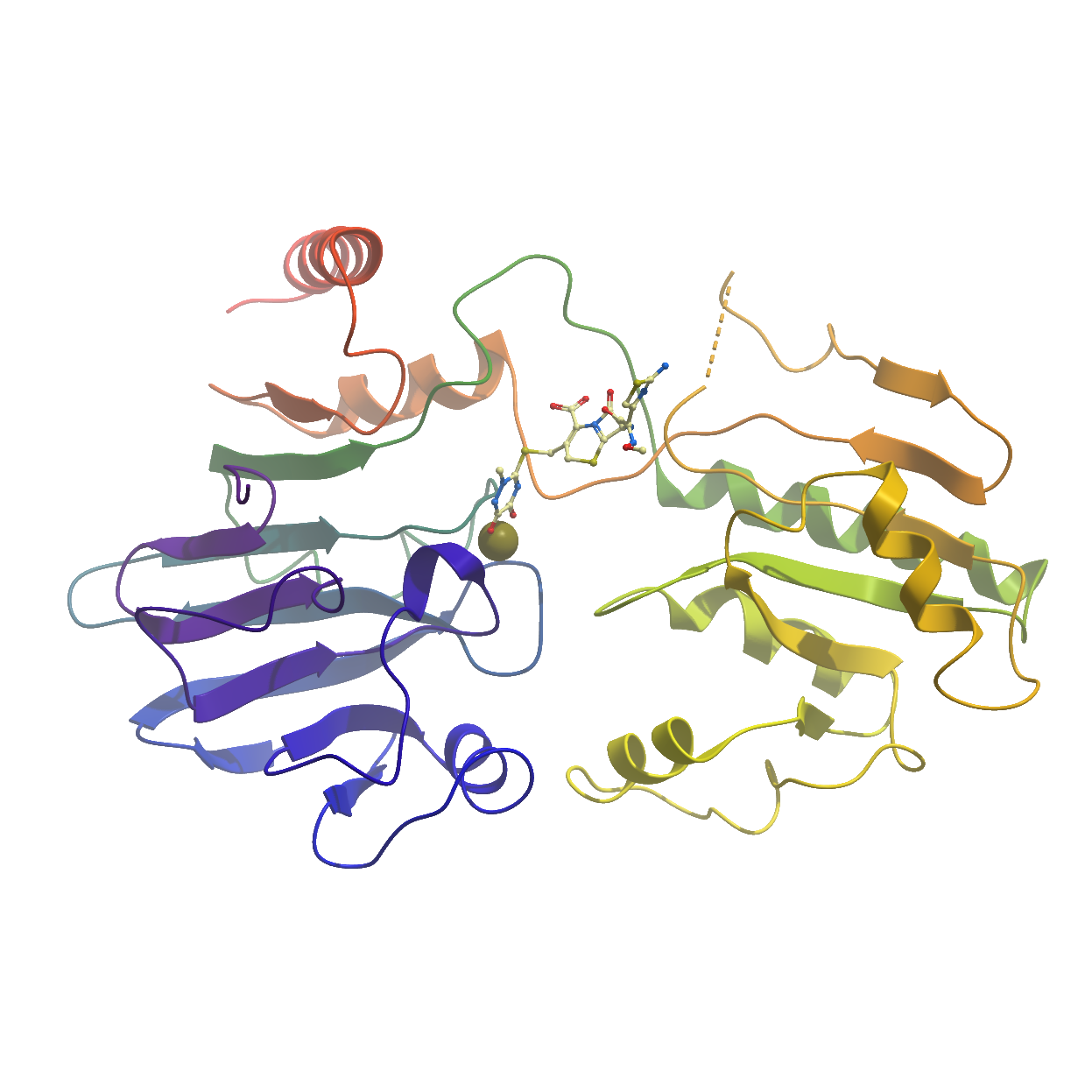
Structure Determination
Crystallization: Protein crystallization was performed by vapour diffusion in sitting drops at 4°. A protein solution at 9-10 mg/mL was mixed at with an equal volume crystallization solution containing 30% PEG 1000, 0.1M MIB pH 6.0 (MIB is Sodium malonate dibasic monohydrate, Imidazole, Boric acid). The crystals were backsoaked overnight in a solution containing 30% PEG 1000, 0.1 M Hepes pH 7.0 to remove the malonate ion from the active site and were subsequently soaked overnight in a solution containing an additional 20 mM ceftriaxone before being loop mounted and plunged directly into a pool of liquid nitrogen.
Data Collection: Data was collected to 2.4Å resolution at ESRF beamline BM30B, and processed using XDS.
Data Processing: The structure was solved by molecular replacement using the program MOLREP and PDBid 5AHO as a search model. Refinement was performed using REFMAC to a final Rfactor = 22.6%, Rfree = 31.5%.