Crystallization, data collection, and structure determination
A DNA fragment encoding the methyltransferase domain of human PRMT4 (residues 140–480) was cloned into a baculovirus expression vector pFBOH-MHL (http://www.thesgc.org/sites/default/files/toronto_vectors/pFBOH-MHL.pdf). The protein was expressed in Sf9 cells as an N-terminal hexa-His tag fusion protein and purified by metal chelating affinity chromatography (TALON resin; Clontech, Mountain View, CA, USA) followed by size-exclusion chromatography (Superdex 200; GE Healthcare). Pooled fractions containing PRMT4 were subjected to tobacco etch virus treatment to remove the His-tag. The protein was purified to homogeneity by ion-exchange chromatography.
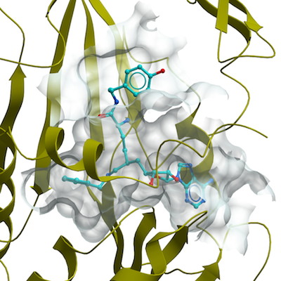
Purified PRMT4 (6.5 mg ml−1) was crystallized with the sitting drop vapor diffusion method at 20 °C by mixing 1 µL of protein solution with 1 µL of the reservoir solution containing 25% PEGG3350, 0.1M ammonium sulfate and 0.1 M Hepes pH7.5. (S)-SKI-72 (0.2 mL of 10 mM in DMSO) was added to the drops with apo crystals and incubated overnight.
X-ray diffraction data for PRMT4 + (S)-SKI-72 were collected at 100 K on a Rigaku FR-E superbright X-ray generator. Data were processed using the HKL-3000 suite [31]. The structure of PRMT4 + TP-064 was isomorphous to PDB entry 4IKP, which was used as a starting model. REFMAC was used for structure refinement. Geometric restraints for compound refinement were prepared with GRADE v.1.102 developed at Global Phasing Ltd. (Cambridge, UK). The COOT graphics program was used for model building and visualization, and MOLPROBITY was used for structure validation.