Molecular Biology
Entry Clone Source: Complex
SGC Construct ID: XX01SUCLG1A-c001
Protein Region: None-None
Vector: pNic-Bsa4
Tag: N-6HIS;N-TEV
Host: BL21(DE3)-R3-pRARE2
Note this plasmid encodes two genes (below) interspersed by a ribosomal binding site.
Sequence (with tag(s)):
SUCLG1A:
MHHHHHHSSGVDLGTENLYFQSMSYTASRQHLYVDKNTKIICQGFTGKQGTFHSQQALEYGTKLVGGTTPGKGGQTHLGLPVFNTVKEAKEQTGATASVIYVPPPFAAAAINEAIEAEIPLVVCITEGIPQQDMVRVKHKLLRQEKTRLIGPNCPGVINPGECKIGIMPGHIHKKGRIGIVSRSGTLTYEAVHQTTQVGLGQSLCVGIGGDPFNGTDFIDCLEIFLNDSATEGIILIGEIGGNAEENAAEFLKQHNSGPNSKPVVSFIAGLTAPPGRRMGHAGAIIAGGKGGAKEKISALQSAGVVVSMSPAQLGTTIYKEFEKRKML
SUCLA2A:
MNLSLHEYMSMELLQEAGVSVPKGYVAKSPDEAYAIAKKLGSKDVVIKAQVLAGGRGKGTFESGLKGGVKIVFSPEEAKAVSSQMIGKKLFTKQTGEKGRICNQVLVCERKYPRREYYFAITMERSFQGPVLIGSSHGGVNIEDVAAESPEAIIKEPIDIEEGIKKEQALQLAQKMGFPPNIVESAAENMVKLYSLFLKYDATMIEINPMVEDSDGAVLCMDAKINFDSNSAYRQKKIFDLQDWTQEDERDKDAAKANLNYIGLDGNIGCLVNGAGLAMATMDIIKLHGGTPANFLDVGGGATVHQVTEAFKLITSDKKVLAILVNIFGGIMRCDVIAQGIVMAVKDLEIKIPVVVRLQGTRVDDAKALIADSGLKILACDDLDEAARMVVKLSEIVTLAKQAHVDVKFQLPIWQ
Protein Expression
Medium: Terrific broth
Antibiotics: Kanamycin, Chloramphenicol
Procedure: Protein was expressed in BL21(DE3)-R3-pRARE2 for 16 hr at 4°C after induction with 200 μM IPTG at an OD of 0.6 measured at 600nm.
Protein Purification
Procedure: Extraction Buffer: 500 mM NaCl, 50 mM HEPES pH 7.5, 20 mM imidazole, 0.5 mM TCEP, 5% glycerol, 1:1000 of Merck Protease Cocktail II.
Binding Buffer: 500 mM NaCl, 50 mM HEPES pH 7.5, 20 mM imidazole, 0.5 mM TCEP, 5% glycerol.
Washing Buffer: 500 mM NaCl, 50 mM HEPES pH 7.5, 40 mM imidazole, 0.5 mM TCEP, 5% glycerol.
Elution Buffer: 500 mM NaCl, 50 mM HEPES pH 7.5, 250 mM imidazole, 0.5 mM TCEP, 5% glycerol.
GF buffer: 500 mM NaCl, 50 mM HEPES pH 7.5, 0.5 mM TCEP, 5% glycerol
A 6L cell pellet was re-suspended in 120ml extraction buffer. Cells were lysed via sonication. The cell debris was removed via centrifugation using a JA 17.00 rotor at 35K for 1hr at 4°C. The supernatant was incubated with 2.5 ml Ni-NTA for 1 hr. Ni-NTA resin bound to the target proteins was washed two times with 50 ml of binding buffer followed by two times with 20 ml of wash buffer and eluted in 5 ml fractions with elution buffer. Elution’s containing protein were loaded onto a S200 gel filtration column and purified via size exclusion. The protein was subsequently incubated with TEV protease at a ratio of 20:1 mg/ml (protein:TEV) overnight at 4°C. Cleaved protein was passed back over 1 ml of Ni-NTA and the flow through containing protein was concentrated to 7 mg/ml.
Columns: Column 1: 2 ml Ni; Column 2: S200 ; Column 3: Ni rebind
Concentration: 7.0 mg/ml
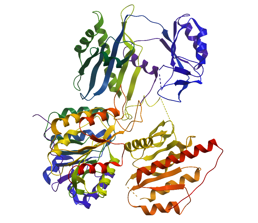
Structure Determination
Crystallization: Crystals were grown by vapour diffusion method. sitting drops containing 100 nL protein (6.2 mg/mL) and 50 nL well solution containing 2.1M DL- malic acid were equilibrated at 20°C.
Data Collection: Beamline: Dmnd I04; Resolution: 2.59 Å
Data Processing: Data was processed using Xia2 auto processing pipeline. The protein crystalised in the trigonal P3121 space group and contained 1 copy of each subunit in the asymmetric unit. The structure was solved via molecular replacement with 1EUC. The final model was produced by iterative cycles of restrained refinement and model building using REFMAC5 and COOT.